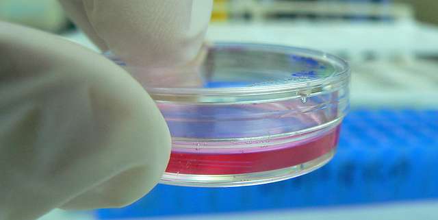
Spectrophotometers measure absorption values and can quantify cell growth in changes both rapidly and effectively in cell culture samples. Image Source: Flickr user Umberto Salvagnin

Cell growth monitoring is required in biomedical research to help determine the rate of change in cell-based tissues. A colorimetric assay is often used to quantify cell growth changes and provides a rapid and precise analysis about the basic health of these cells and can monitor their response to medications. This information is important for measuring metabolic activity, bacterial growth in cell populations, and can provide DNA analysis. Colorimetric assay data can then be used to develop treatment plans and monitor their success, providing a valuable resource in the medical research field.

Spectrophotometers provide specific cell information which is used to develop a colorimetric assay that can measure a variety of cell characteristics. This method of analysis is highly sensitive and can accurately quantify up to 3000 cells per petri dish1 and analyze their functioning and changes with UV and visible spectral absorption value measurements. Simple to use, non-destructive, with rapid and repeatable results, spectrophotometers are an essential tool in the medical science and research technologies of the future.

READ  Active Pharmaceutical Ingredient Detection Using Spectrophotometers: An Investment that Pays Off

Cell proliferation assays

A cell-proliferation assay2 can monitor various parameters of cell growth and functioning. Spectral analysis is often used to develop a colorimetric assay for cell-proliferation that can determine cell viability and toxicity when exposed to various factors of conditions. A colorimetric assay can be used for quantification proliferation and profiling, for disease pattern identification and treatment monitoring analysis.

Sample requirements to develop a cell proliferation or colorimetric assay require only simple and affordable reagents for color absorption monitoring. With these reagents, cell structure, growth, and changes can all be monitored effectively. This data can then be used to monitor cell viability and provide information in relation to large tissue analysis.

DNA analysis and metabolic activity

One common method for assessing cell proliferation is to develop a colorimetric assay based on DNA content in cells. According to the Biocompare online article Common Methods of Measuring Cell Proliferation, “new DNA synthesis assays provide a precise marker of proliferation at the population level or the individual cell level [and] can be multiplexed with other cellular markers like mitochondrial function or cell morphology.” These new developments in technology open the door to many new research opportunities and medical treatment monitoring options. A colorimetric assay can provide a wealth of information about DNA content and offers a way to quantify these changes at various given stages to create comparison models.

13107953_463b39146b_o.jpg

Colorimetric assays can be used to quantify DNA in mammalian cell structures. Image Source: Flickr user Rob Pongsajapan

Metabolic activity can also be measured using spectrophotometric technology by monitoring the activity of cells when exposed to tetrazolium salts3 and quantifying the color changes in relation to this activity. This method of analysis can be used to perform multiple assays and can also be used with other color indicators or reactive additives.

INFO  Get More Information

Bacterial cell division

A colorimetric assay can also be used to analyze and quantify cell division. Bacterial cells divide quickly and can be monitored using a culture sample. Monitoring the growth and population rate increase of bacteria and help identify specific properties of bacteria which can indicate maturity and regulate treatment options.

E. coli, a common yet dangerous form of bacteria shows growth in three distinct stages. Developing a colorimetric assay of an E. coli bacterial sample4 can be used to estimate bacterial population and rate of increase. Spectrophotometers can measure the turbidity (cloudiness) of a culture and monitor its rate of change to quantify bacterial counts.

Colorimetric assays are commonly used for bacterial analysis and can be used to estimate cell growth and contamination rate. Image Source: Flickr user SonoranDesertNPS

Other microbial applications of spectrophotometry have been used to detect bacterial contamination in pharmaceutical formulations and other bacterial inhibiting substances. The ability to detect and monitor various bacterium, quantify these levels, and predict growth rates have been invaluable to both the pharmaceutical and biomedical sciences.

Applications of spectrophotometry in medicine

Using a colorimetric assay and spectrophotometric technology have opened the door to new applications in medical science and research capabilities. Spectrophotometers offer an affordable and efficient method of analysis for many various applications in these fields. As technology continues to grow, instrumentation options and availability continues to grow as well. At HunterLab we specialize in spectrophotometric application in pharmaceutical and medical technology. We continually work with industry leaders to develop instrumentation to meet the current needs of the healthcare and medical science industries. For more information on the various applications of spectrophotometry in these areas, contact HunterLab today.